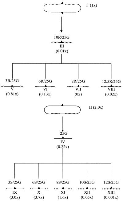
Figure 2.
Flow diagram for the generation of modified single-stranded oligonucleotides. The upper targeting strands of chimeric oligonucleotides I and II are separated into pathways resulting in the generation of single-stranded oligonucleotides that contain (A) 2′-O-methyl RNA nucleotides or (B) phosphorothioate linkages. Fold changes in repair activity for correction of kans in the HUH7 cell-free extract are presented in parentheses. Each single-stranded oligonucleotide is 25 bases in length and contains a G residue mismatched to the complementary sequence of the kans gene. The numbers 3, 6, 8, 10, 12 and 12.5 indicate how many phosphorothioate linkages (S) or 2′-O-methyl RNA nucleotides (R) are at each end of the molecule. Hence oligo 12S/25G contains an all phosphorothioate backbone, displayed as a dotted line. Smooth lines indicate DNA residues, wavy lines indicate 2′-O-methyl RNA residues and the carat indicates the mismatched base site (G).

