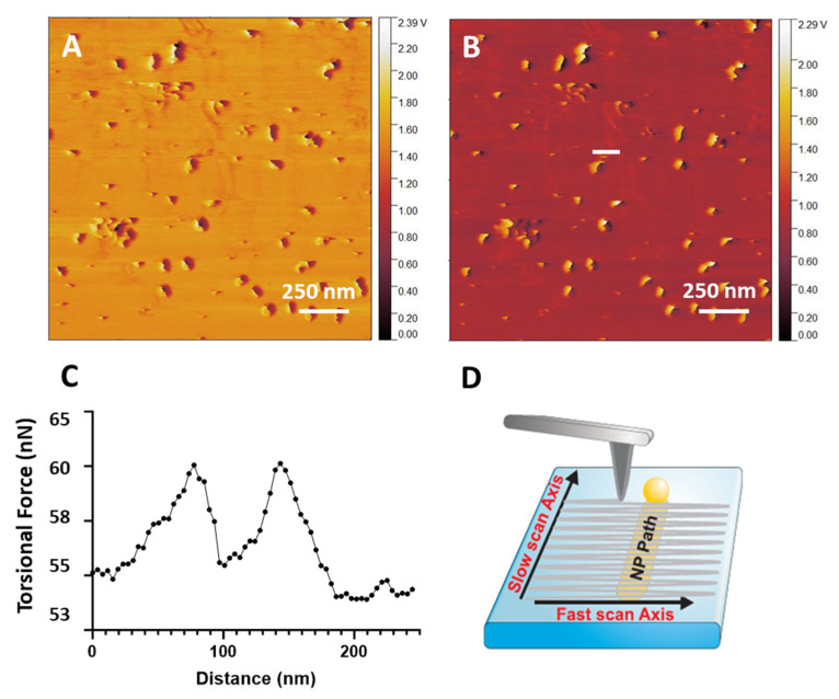
Figure 10.
(A) LFM lateral deflection trace and (B) retrace images showing the two-axis movement of AuNPs on MePPOx over a 2 × 2 µm scan with a Set Point of 40 nN. The white line highlights the location where the scan was conducted. (C) A 2D plot across the ‘drag lines’ in (A), and (D) illustration of the tip’s raster scan motion moving a AuNP in the slow axis.

