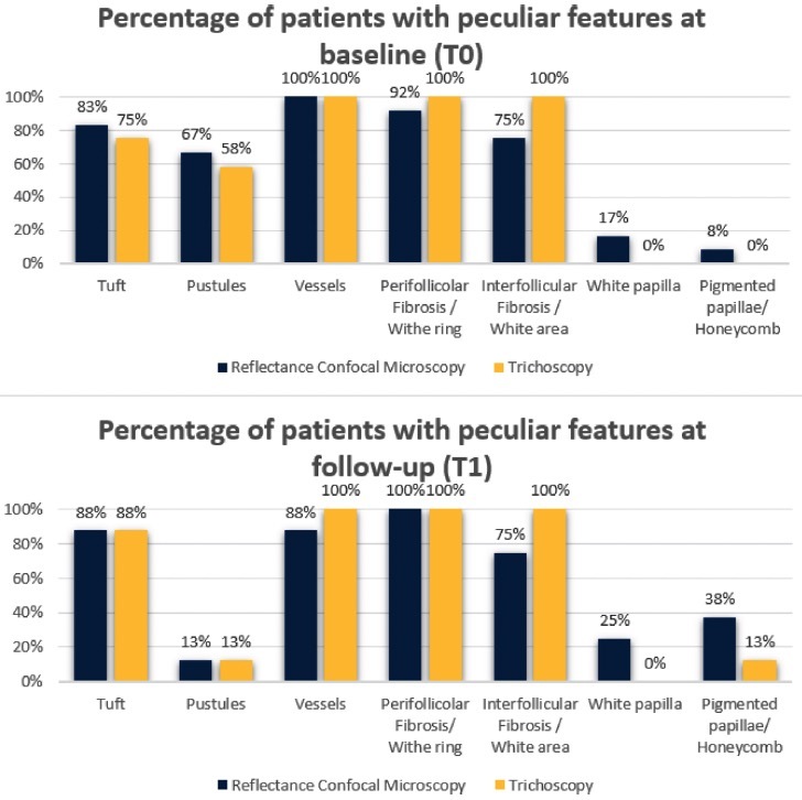
Figure 2.

Results of reflectance confocal microscopy (RCM) features of folliculitis decalvans and the corresponding features visualized by trichoscopy at baseline (T0) and at follow-up after three months of therapy (T1). The perifollicular fibrosis seen with RCM matched the white rings visualized by trichoscopy. Similarly, the interfollicular fibrosis detected by RCM was aligned with the white areas found by trichoscopy. The pigmented papillae evident in RCM analysis corresponded to the honeycomb patterns identified by trichoscopy. However, the white papillae identified by RCM did not have a trichoscopic counterpart.
