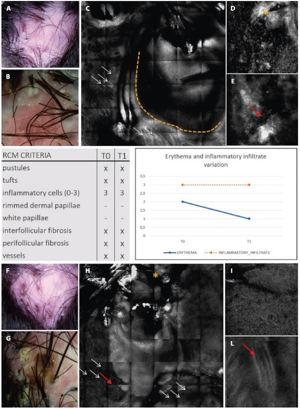
Figure 5.

Clinical, trichoscopic and confocal aspects of patient 2 at (A–E) T0 and (F–I) after treatment at T1. (A,B) This patient presents on trichoscopy a decrease of erythema score to T0 and T1, while on reflectance confocal microscopy (RCM) inflammatory cells are still present as sign of disease activity. Clinical and trichoscopic appearance at T0. (C) Follicular pustule (yellow dashes) and perifollicular fibrosis (white arrows) seen with RCM. (D,E) RCM shows inter follicular fibrosis (yellow asterisk) and superficial dilated vessels (red arrow). (F,G) Clinical and trichoscopic features of active FD at T1. (H,I) Confocal magnification demonstrates the presence of numerous inflammatory cells (white arrows), interfollicular fibrosis (yellow asterisk), and dilated superficial vessels (red arrows).
