
Figure 1.
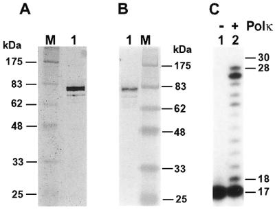
Analyses of purified human Polκ. (A) Purified human Polκ (200 ng) was analyzed by electrophoresis on a 10% SDS–polyacrylamide gel and visualized by silver staining. Protein size markers (lane M) are indicated on the left. (B) Purified human Polκ (40 ng) was analyzed by a western blot using a mouse monoclonal antibody against the His6 tag. Protein size markers (lane M) are indicated on the right. (C) DNA polymerase assays were performed without (lane 1) or with (lane 2) purified human Polκ (0.2 ng, 2 fmol), using the 30mer DNA template, 5′-CCTTCTTCATTGGAACATACTTCTTCTTCC-3′, annealed with the 5′-32P-labeled primer, 5′-GGAAGAAGAAGTATGTT-3′. DNA size markers in nucleotides are indicated on the right.