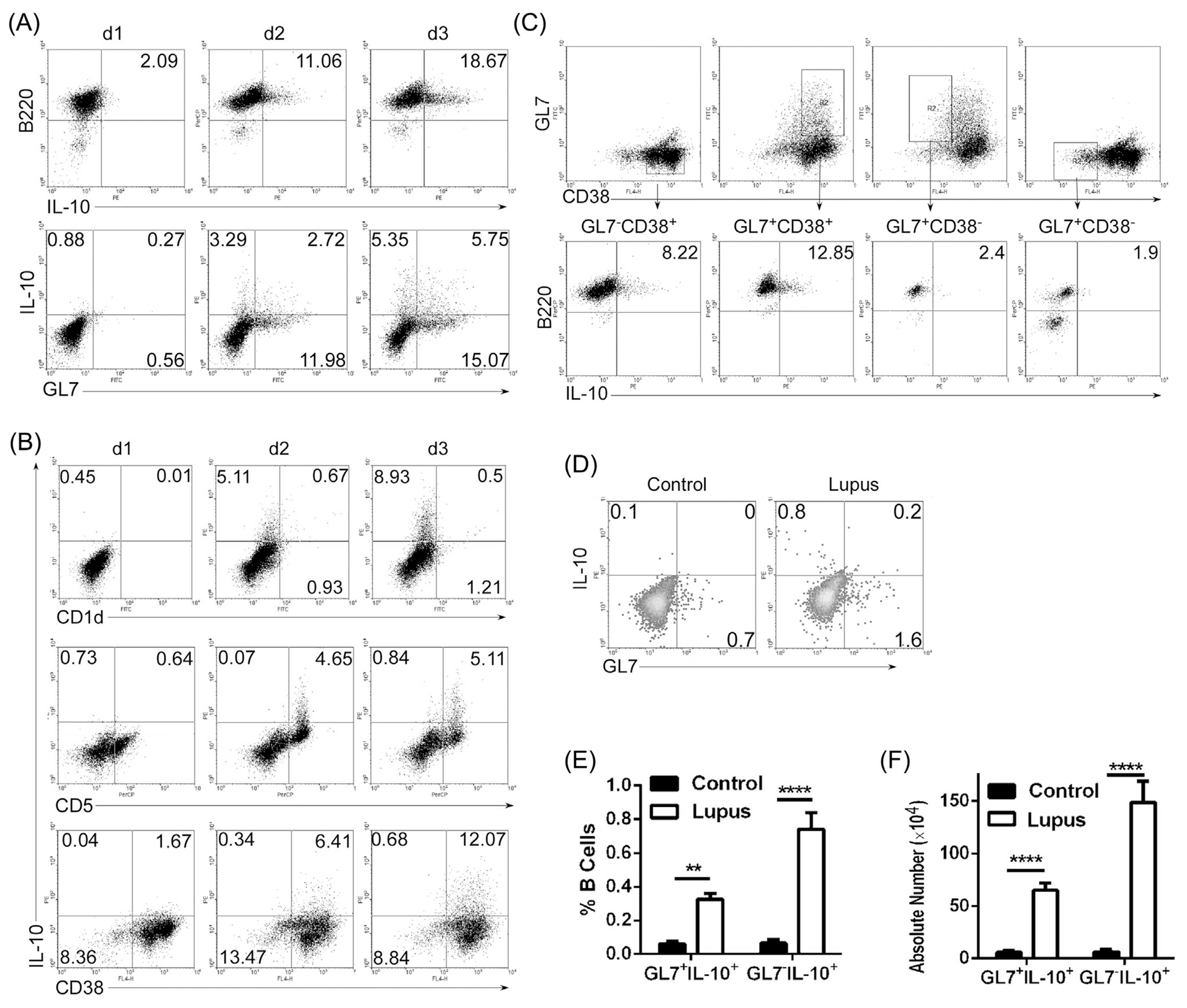
Fig 1.

GL7−IL-10+ B cells are expanded in LPS-stimulated B cells and spleen of mice with lupus-like disease. (A–C) Splenic B cells were stimulated for 1–3 days with LPS (1 μg/ml) and analyzed by the intracellular cytokine-staining assay. Cells were gated for B220+ or GL7+ B cells (A, C) or CD1d+, CD5+, CD38+ B cells (B) and quadrants indicate percentage of IL-10-expressing cells. (D–F) Splenic lymphocytes from lupus-like MRL/lpr (Lupus) mice were analyzed by the intracellular cytokine-staining assay. Quadrants indicate percentages of IL-10-expressing GL7− or GL7+ B cells. Statistical analysis of the percentages (E) or absolute number (F) of IL-10-expressing GL7− or GL7+ B cells. Data represent at least four independent experiments. (E, F) Data were analyzed by Student’s t-test (two tailed). Error bars, s.e.m. **P < 0.01, ****P < 0.0001.
