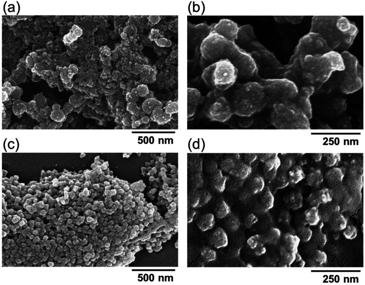
FIG. 3.
Morphology and microstructure of calcium phosphate nanoparticles analyzed via SEM. (a) Bare calcium phosphate nanoparticles were observed under 60 000× magnification and (b) under 140 000× magnification. (c) pDNA-CaP NPs were observed under 60 000× magnification and (d) under 140 000× magnification.

