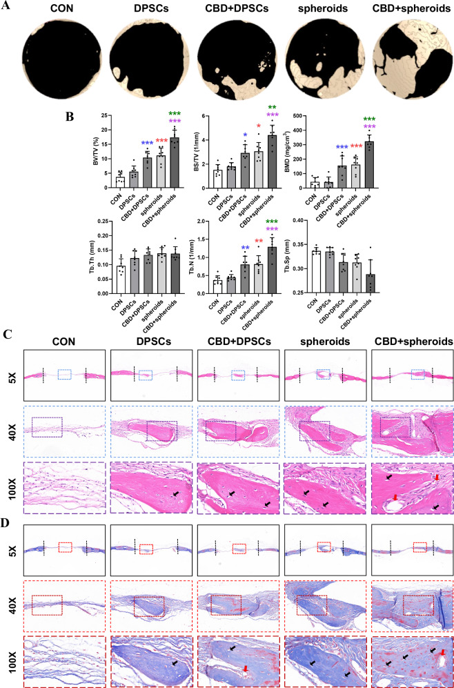
Fig. 6. CBD-treated microspheroids promoted bone regeneration in mice calvarial bone defects.
A Micro-CT images of calvarial bone defects after 8 weeks. B Quantitative analysis of newly formed bone parameters, bone volume/total volume (BV/TV), bone surface/total volume (BS/TV), bone mineral density (BMD), trabecular thickness (Tb.Th), trabecular number (Tb.N) and trabecular separation (Tb.Sp) (n = 8). Representative microscopic images of histological tissue sections: C HE staining and D Masson staining. Osteoblast (yellow arrows); Vessel (red arrows). Significant difference compared different groups, *P < 0.05, **P < 0.01, and ***P < 0.001; compared between DPSC and CBD+DPSC groups in blue; compared between DPSC and spheroids groups in red; compared between spheroids and CBD+spheroids groups in green; compared between CBD+DPSC and CBD+spheroids groups in purple.

