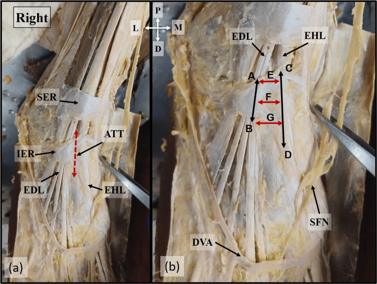
Figure 1. Location of ATT.
Location of the ATT in (a) and the dimensions of the ATT in (b). A-B: Lateral boundary. C-D: Medial boundary. E, F, and G denote the width of ATT at the proximal, middle, and distal ends
ATT: anterior tarsal tunnel; EHL: extensor hallucis longus; EDL: extensor digitorum longus; SER: superior extensor retinaculum; DVA: dorsal venous arch; SFN: superficial fibular nerve; P: proximal; D: distal; M: medial; L: lateral

