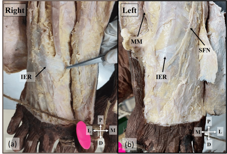
Figure 2. Absence of ATT.
Absence of the ATT due to the displaced IER distally from its normal position on the right (a) and left side (b) of the 60-year-old male cadaver
ATT: anterior tarsal tunnel; IER: inferior extensor retinaculum; MM: medial malleolus; SFN: superficial fibular nerve; P: proximal; D: distal; M: medial; L: lateral

