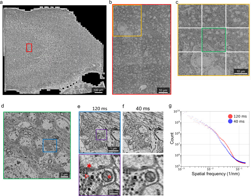
Fig. 3. High-throughput millimeter-area imaging with beam deflection.
a A fully stitched montage of a hippocampus section (1 mm2) imaged with a Cricket-equipped TEM, at a pixel size of 3 nm. The montage includes 4320 tiles (or 480 supertiles). Representative data of over 3000 sections. b A montage of 6 stitched supertiles (from red outline in a). c A Cricket supertile consisting of 9 tiles (from orange outline in b). Each tile has 6000 × 6000 pixels and the overlap between neighboring subtiles of a supertile is 15% (~900 pixels). d A single image tile (from green outline in c). e Further zoom-in shows postsynaptic density (*), synaptic vesicles (△) and a cross-section plane of a mitochondria (☆). f Same area and zoom-in as in e but imaged with 40 ms exposure time. The whole section is imaged with 120 ms exposure time a–e. g SNRs of the images (e, f) are computed with the same procedure as Fig. 2d. Scale bars, 100 μm a, 10 μm b, c, 2 μm d, 1 μm (e, upper), 200 nm (e, lower).

