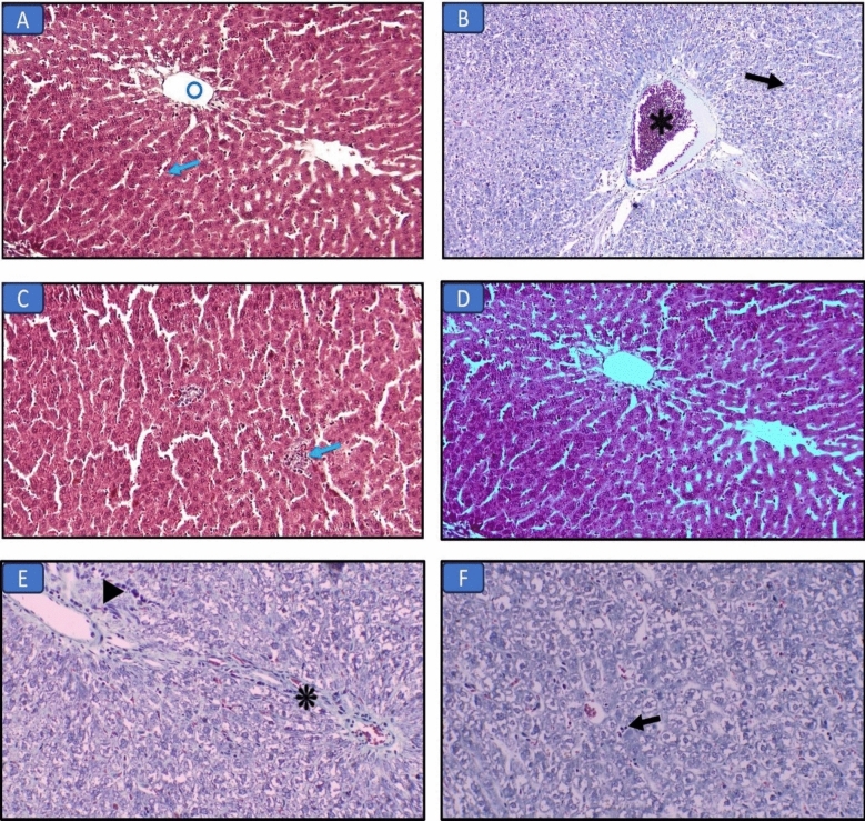
Figure 2.
Masson’s trichrome-stained liver sections in the different groups: (A) Control group, normal liver structure with central vein (circle) and hepatocytes (arrow) (×200). (B) Long chain saturated fat diet group, the liver shows diffuse injury with necrosis (arrow) and congestion (asterisk) (×200). (C) Long chain monounsaturated fat diet group, the liver shows focal mild lymphocytic infiltrate (arrow) (×200). (D) Long chain polyunsaturated fat diet group, the liver structure was normal regarding hepatocytes and bile canaliculi (×200). (E) Medium chain fat diet group, the liver shows fibrous tissue band (asterisk) and focal mild lymphocytic infiltrate (arrowhead) (×200). (F) Short chain fat diet group, the liver shows focal mild lymphocytic infiltrate (arrow) (×200).

