Figure 1.
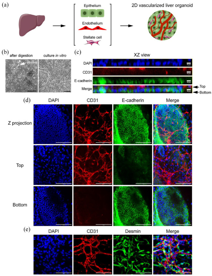
2D vascularized liver organoids. (a) Workflow illustration. (b) The phase contrast images of the primary liver cells right after digestion and those cultured in vitro in regular dishes for 4 days. The white arrow pointed to a microvessel. (c–e) Immunostaining of the 2D vascularized liver organoids cultured for 4 days in vitro. The antibodies were against CD31, E-cadherin, and Desmin. DAPI stained nuclei. Scale bars, 100 µm.

