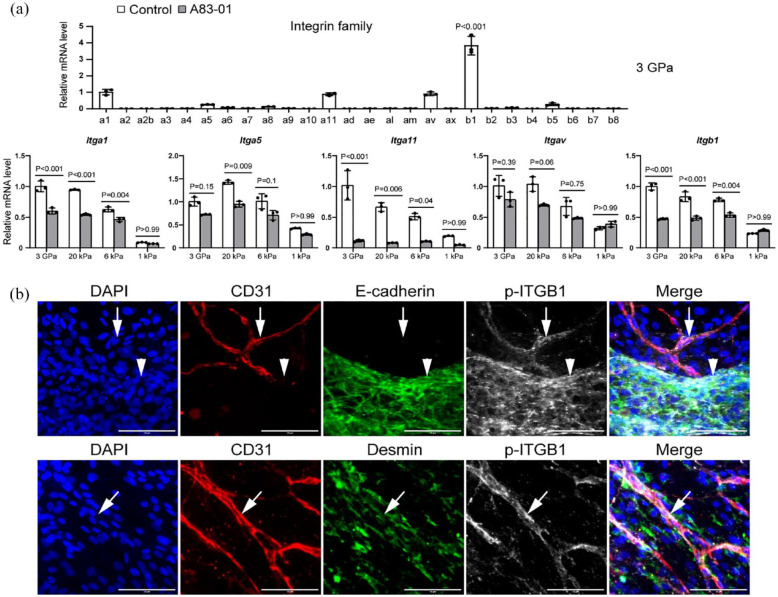
Figure 6.
Integrins in vascularized liver organoids. (a) The qPCR analysis of vascularized liver organoids on the ECM of different stiffness. N = 3. One-way or two-way ANOVA was performed on the data, followed by Bonferroni post hoc tests. p < 0.05 was considered significant. (b) Immunostaining of vascularized liver organoids cultured on 3 GPa ECM in the control medium by the antibodies against CD31, E-cadherin, and p-ITGB1 (Y783). DAPI stained nuclei. Arrows pointed to CD31+ p-ITGB1+ microvascular endothelial cells. Arrowheads pointed to E-cadherin+ p-ITGB1+ epithelial cells. Scale bars, 100 µm. The cell culture time was 1 week.

