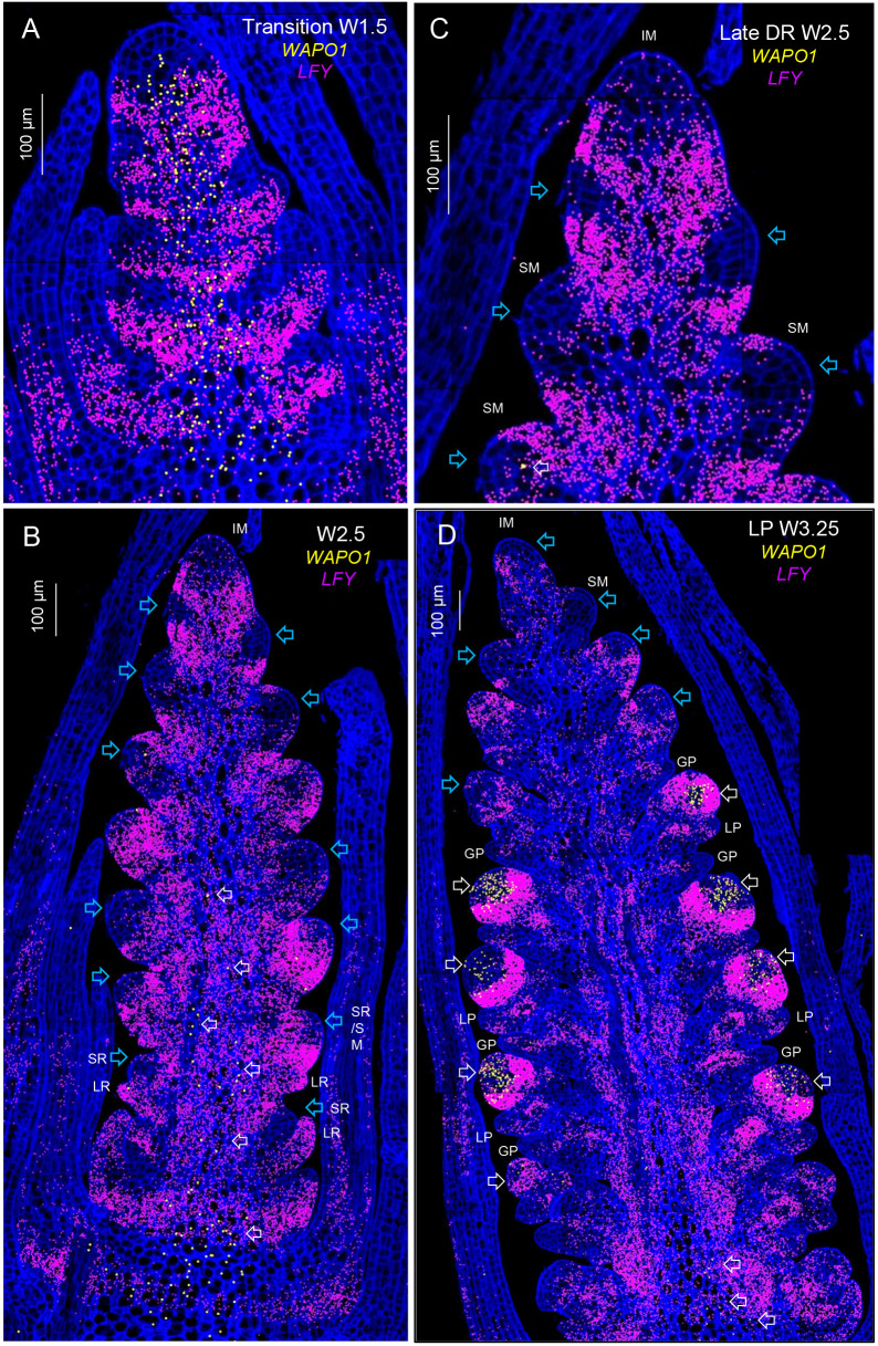
Fig. 4.
Single-molecule fluorescence in-situ hybridization (smFISH) of LFY and WAPO1 during spike development. Cell walls stained with calcofluor are presented in dark blue. (A) Elongated shoot apical meristem transitioning from a vegetative to an inflorescence meristem (IM, W1.5). (B) Late double-ridge stage (W2.5). (C) Detail of the IM region from B. (D) Lemma primordia stage (W3.25). Blue arrows indicate regions of the SM where LFY expression is lower and white arrows show regions of WAPO1 expression. GP, glume primordium; LP, lemma primordium; LR, leaf ridge; SR, spikelet ridge. Scale bars: 100 μm. W, Waddington scale (Waddington et al., 1983).

