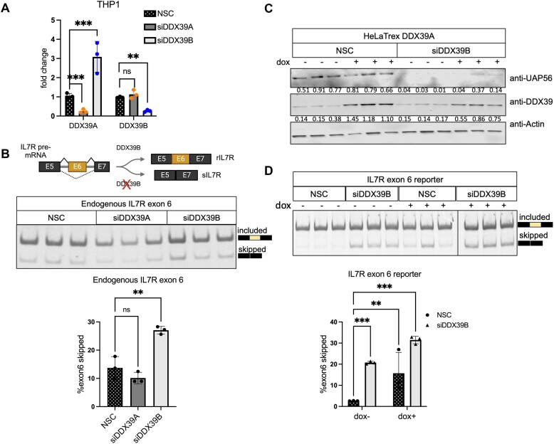
Figure 4.
DDX39A does not regulate IL7R exon 6 splicing. (A) RT-qPCR analysis of the transcript levels of DDX39A and DDX39B upon DDX39A knockdown (siD09) and DDX39B knockdown (siD13) compared to control (NSC) in THP1 cells. Statistical significance was assessed using Student's t test (***P ≤ 0.001; **P ≤ 0.01; ns = not significant). (B) The top panel provides a diagrammatic representation of IL7R exon 6 splicing in the presence and absence of DDX39B. The bottom panel shows the RT-PCR analysis of IL7R exon 6 splicing in transcripts from endogenous IL7R. (C, D) Rescue experiments of IL7R exon 6 splicing in HeLa cells stably expressing siRNA-resistant DDX39A transgene under the control of doxycycline promoter. (C) Immunoblot depicting the protein abundance of DDX39A and DDX39B with respect to actin. The numbers indicating protein abundance relative to actin are listed below each immunoblot. (D) RT-PCR analysis of IL7R exon 6 splicing from minigene reporter. In all panels, the data are shown as mean ± s.d., and statistical significance was assessed using one-way ANOVA (****P ≤ ***P ≤ 0.001; **P ≤ 0.01; ns = not significant).

