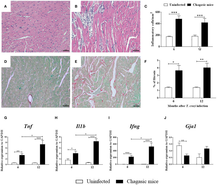
Figure 2.
Morphological analysis and gene expression of pro-inflammatory cytokines in the hearts of uninfected and chagasic mice at 6 and 12 months after infection. (A, B) Representative micrographs of hematoxylin and eosin-stained heart sections of uninfected and chagasic mice at 12 months following infection. (C) Inflammatory cells quantified by morphometric analysis. (D, E) Micrographs of picrosirius red-stained heart sections of uninfected and chagasic mice. (F) Fibrotic area represented by percentage of collagen deposition in heart sections. Gene expression of pro-inflammatory cytokines (G) Tnf, (H) Il1b, (I) Ifng, and (J) Gja1 assessed by RT-qPCR using cDNA samples prepared from mRNA extracted from experimental mouse hearts. Values represent means ± S.E.M. of 5-6 mice per group. ***P< 0.001; **P< 0.01; *P<0.05 compared to uninfected group.

