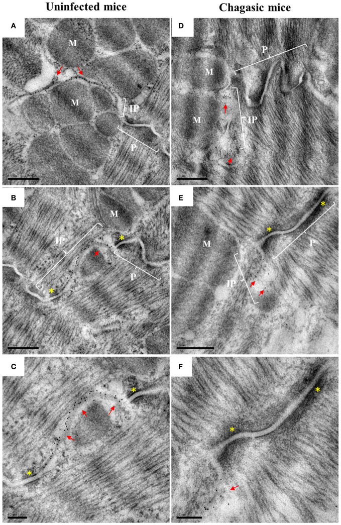
Figure 5.
Ultrastructural analysis of total Cx43 in intercalated discs. Hearts from uninfected and chagasic control mice (n = 3 per group) were processed for analysis by transmission electron microscopy. Ultrathin sections were incubated with total anti-Cx43 antibody, with labeling revealed by the immunogold technique. (A–C) representative sections from uninfected control mice; (D–F) representative sections from chagasic mice. Red arrows indicate the presence of gap junctions in the intercalated discs; yellow asterisks indicate desmosome; P, plicate; IP, interplicate; M, myofibrils. Scale bars = 0.5 µm (A, B, D, E), 0.2 µm (C, F).

