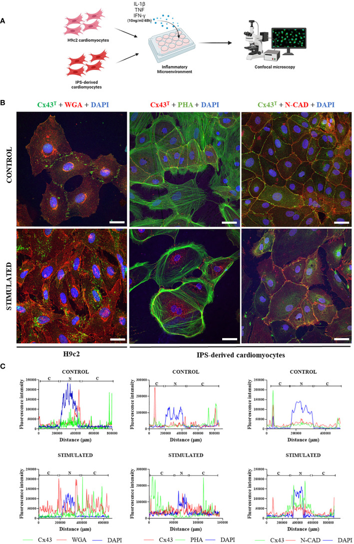
Figure 7.
Effect of inflammatory microenvironment on Cx43 distribution in vitro. (A) Experimental in vitro design involving H9c2 cells and iPSC-derived cardiomyocytes, with both cell types stimulated by pro-inflammatory cytokines (IL-1β, TNF, and IFN-γ, 10 ng/ml of each cytokine) for 48 hours for immunofluorescence analysis. (B) Immunofluorescence of H9c2 cells and iPSC-derived cardiomyocytes; (C) Analysis of Cx43 distribution by fluorescence intensity in H9c2 cells and iPSC-derived cardiomyocytes after 48 hours of stimulation with pro-inflammatory cytokines IL-1β, TNF, and IFN-γ. Cells were stained with WGA (red), cell nuclei with DAPI (blue), and total Cx43 with anti-Cx43 antibody (green). Images analyzed by confocal microscopy. Scale bars = 50 µm. WGA, Wheat Germ Agglutinin; Cx43, Connexin 43; IL-1β, Interleukin 1 beta; TNF, Tumor necrosis factor; IFN-γ, Interferon gamma; C, Cytoplasm; N, Cell Nucleus. Created with BioRender.com.

