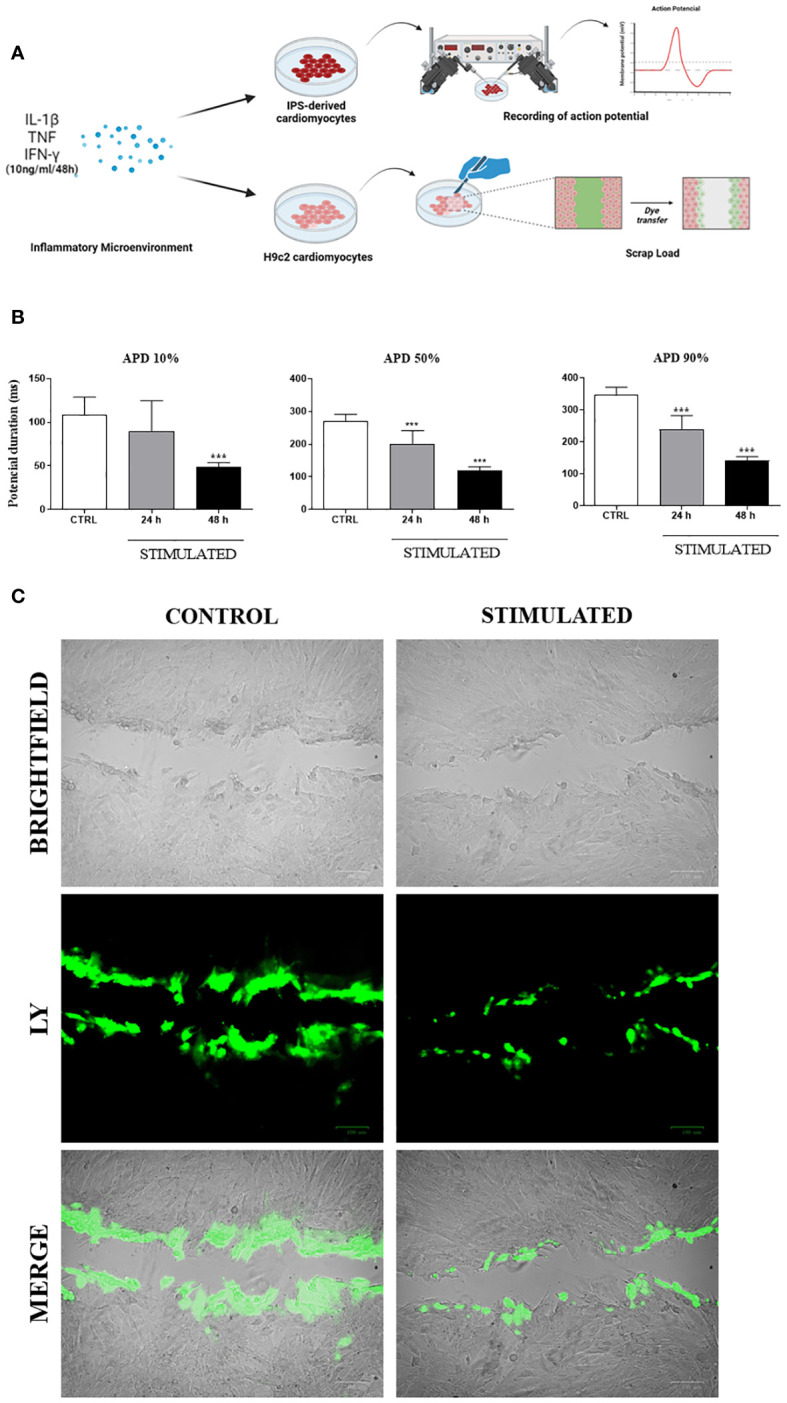
Figure 8.

Functional in vitro analysis of Cx43: Action Potential Duration and Lucifer Yellow dye transfer. (A) Schematic drawing of functional testing performed on iPSC-derived cardiomyocytes and H9c2 cardiomyocytes, stimulated with pro-inflammatory cytokines to respectively analyze the duration of action potential and dye transfer between neighboring cells. (B) After stimulation, action potential duration (APD) of iPSC-derived cardiomyocytes was determined at 10%, 50% and 90% levels. (C) Lucifer Yellow dye transfer test (green) performed in H9c2 cells (bright field) stimulated with pro-inflammatory cytokines IL-1β, TNF, and IFN-γ for 48 hours. Predominance of dye observed in cells at the margin where scalpel incision was made, while absence of stimulation resulted in dye reaching more distant cells; IL-1β = Interleukin 1 beta; TNF = Tumor necrosis factor; IFN-γ = Interferon gamma; CTRL = Control group; ***P< 0.001 compared to unstimulated cells. Scale bars = 100 μm.
