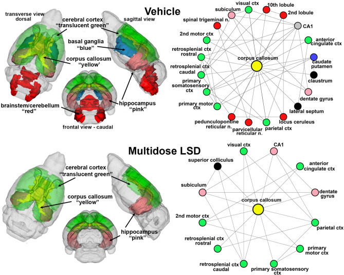
Figure 4.
White matter connectivity. The radial connectivity diagrams show the connections (lines) to different nodes (colored circles) from the corpus callosum (yellow) comparing vehicle to multidose LSD. Circles in green highlight brain areas comprising the cerebral cortex. Circles in red are brain areas located in the brainstem and cerebellum while those in pink are hippocampal brain areas. Area in black are not associated with these other brain regions. The color-coded 3D representation shows the location of these brain areas providing a visual summary of their difference between experimental groups and location.

