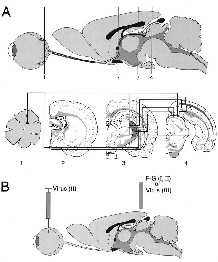
FIG. 1.
Visual nuclear neuronal circuitry and experimental design. (A) Sagittal image of a rat eye, optic nerve, and brain with coronal positions (levels 1 to 4) through the retina and major visual centers illustrated. Axons of retinal ganglion cells (level 1) project, via the optic nerve, to terminate in subcortical visual nuclei of the rat brain including the SCN (level 2), LGN (level 3), and superior colliculus (level 4), also known as the optic tectum. Visual cortical areas (level 4) of the occipital cortex are innervated by projections from subcortical visual centers. In coronal sections, visual nuclei are indicated by areas of darkened shading. Retinal axon projections and interconnections between central visual nuclei are indicated by arrowed lines. Lines with arrowheads at both ends represent commissural connections. For illustration clarity, retinal axon terminal arbors are shown as branches of a single fiber. However, this is not meant to imply that all visual nuclei are innervated by branches of retinocollicular axons. Retinal projection is contralateral to retinorecipient nuclei; interconnections between visual nuclei are ipsilateral. The coronal templates used in this figure were modified from reference 28. In the rat eye infection paradigm, PRV strains expressing gI and gE can spread to and infect central visual projection fields located in the forebrain, such as the hypothalamus (SCN) and the thalamus (LGN, including the dorsal [dLGN] and ventral [vLGN] nuclei, as well as the IGL), in addition to a midbrain target, the superior colliculus. Viruses lacking gI, gE, or both are able to infect only a subset of these retinorecipient regions, including the SCN and the IGL but not the dLGN or superior colliculus. (B) A colocalization experiment was used to examine whether a gI/gE-deleted PRV strain was able to infect retinal ganglion cells which project to the superior colliculus. Superior colliculus-projecting retinal ganglion neurons were identified by stereotaxic injection of the fluorescent tracer F-G into the left superior colliculus of the rat brain (experiment I; F-G alone). Following retrograde axonal transport within retinotectal projections, F-G tracer accumulates in ganglionic neuron cell bodies and processes in the contralateral retina. Intravitreal inoculation of PRV results in viral infection of retinal ganglion neurons. Colocalization of viral antigen and F-G tracer would identify retinotectal projecting neurons as permissive to viral infection (experiment II; F-G and virus). In separate independent experiments, individual PRV strains were inoculated into the left superior colliculus by stereotaxy and virus infection of central visual nuclei and the retina were examined (experiment III; virus alone).

