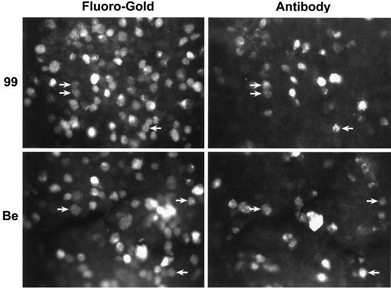
FIG. 3.
Colocalization of virus and tracer in retinotectally projecting retinal ganglion neurons. F-G was injected into the left superior colliculus; this was followed by intravitreal inoculation of either PRV 99 (gI− gE−) or PRV Be (gI+ gE+) into the contralateral (right) eye. F-G inoculation preceded PRV Be injection by 48 h; F-G and PRV 99 inoculations were concurrent. At maximal postinfection survival times (PRV 99, 96 h; PRV Be, 48 h), the animals were sacrificed and their retinas were isolated. Retinal tissue was reacted with rabbit polyclonal antibody Rb133 and TRITC-conjugated secondary antibody. Immunoreacted retinas were whole mounted and visualized by fluorescence microscopy. Each panel doublet (F-G or antibody) represents the same microscopic field. For each virus, results from one representative animal are presented. Retinal ganglion neurons displaying colocalization of viral antigen and tracer are indicated by arrows.

