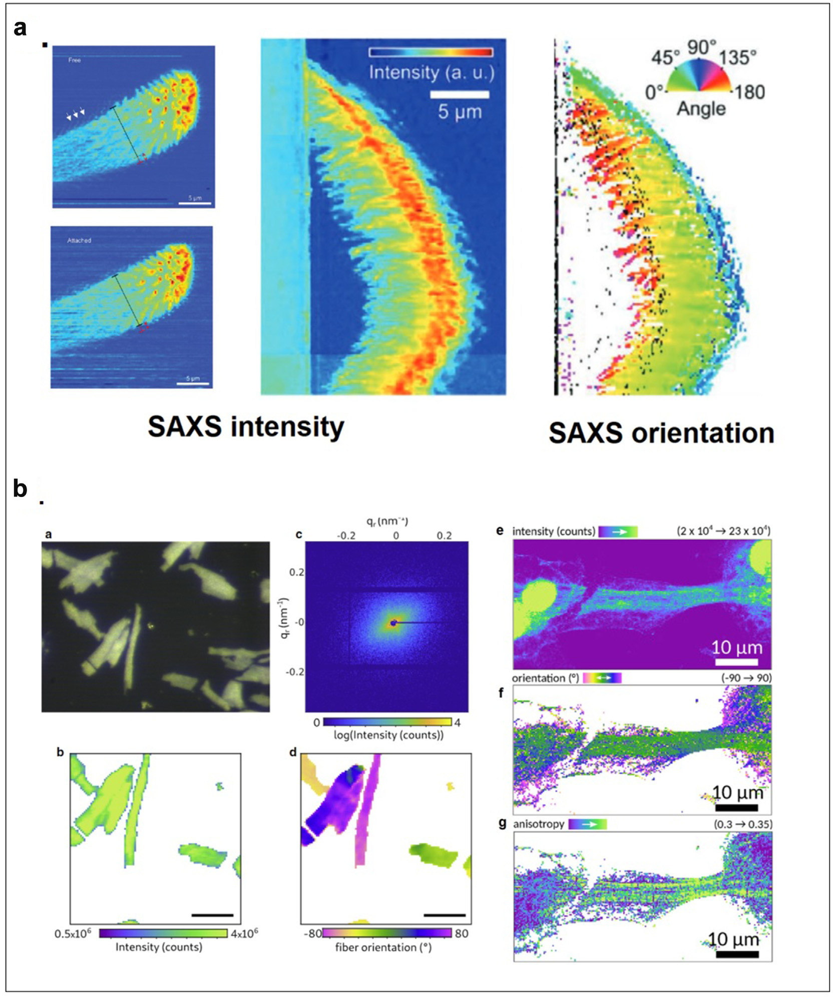
Figure 2.

(a) Physical basis of adhesion by spider attachment hairs. Top view of attachment hair freestanding and attached (right) as visualized by WAXS intensities. Side view of attached hair (middle) and the distribution of orientations in attachment fibrils as determined from SAXS intensity and orientation (Flenner et al., 2020). (b) Structural organization of cardiomyocytes (a) Optical micrograph of a freeze-dried cardiomyocyte recorded with the beamline on-axis microscope (brightfield). (b) X-ray darkfield image from the integrated signal. (c) Diffraction pattern from one scan point. (d) Orientation of constituent actomyosin fibrils is determined by the orientation of diffraction. Scale bars for (a), (b), and (d) 50 mm. (a-d from Reichardt et al., 2020). (e) X-ray darkfield map of an iPS-derived cardiomyocyte. (f) Orientation of actomyosin fibers obtained by PCA analysis. The anisotropy (g) contrasts the highly oriented actomyosin filaments with modulation originating from the striated actomyosin filaments (e-g from Nicolas et al., 2019).
