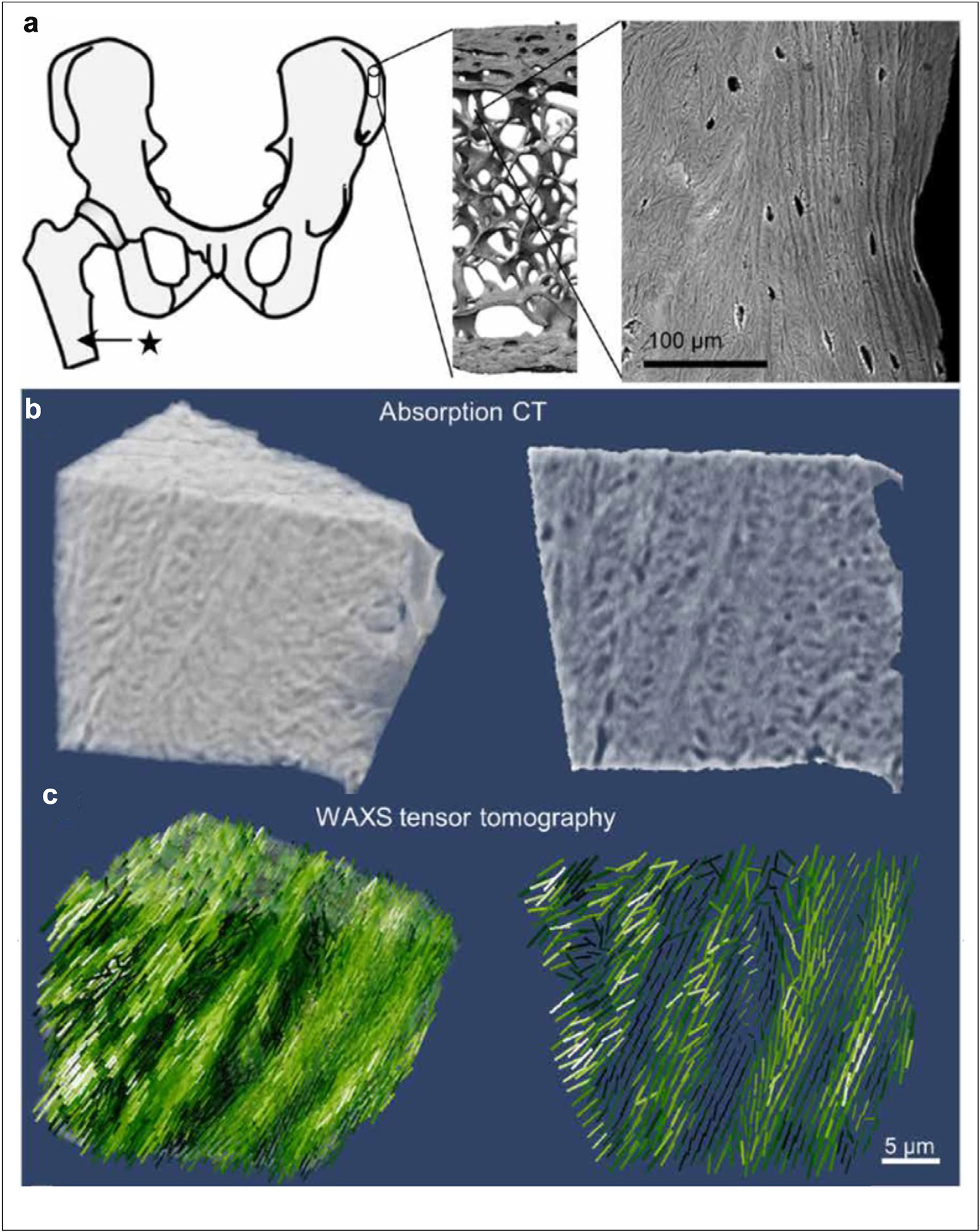
Figure 4.

Mapping the 3D orientation of nanostructures in human bone (a) anatomical location of the sampling site of the bone cube, its hierarchical structure from laboratory microcomputed tomography (microCT) and scanning electron microscopy (SEM). (b) High-resolution absorption tomogram (left) with the two-dimensional image (right) of a central section. (c) Three-dimensional distribution of orientations reconstructed from the WAXS tensor.
