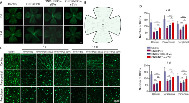
Figure 3.

RGC survival of ONC mice with or without sEV treatment.
(A) Flat-mounted retinas of ONC mice with or without treatment with sEVs on day 7 and day 14 post-ONC. RGC degeneration was progressive after ONC. (B) Schematic diagram of different regions of flat-mounted retinas (central, paracentral, and peripheral). Created with Microsoft PowerPoint Professional 2019. (C) Representative image of central, paracentral, and peripheral retinal regions in ONC model mice with or without treatment with sEVs. Following treatment with hiPSC-sEVs, more RGCs survived in retinas on day 7 post-ONC. However, following treatment with NPC-sEVs, more RGCs survived in retinas on both days 7 and 14. Scale bars: 100 μm. (D) Quantification of mean number of RGCs in different retinal regions at 7 and 14 days (n = 8/group). Data are expressed as mean ± SD. *P < 0.05, **P < 0.01, ***P < 0.001 (one-way analysis of variance followed by Tukey’s post hoc test). hiPSC: Human induced pluripotent stem cell; iPSC: induced pluripotent stem cell; NPC: neural progenitor cell; ns: not significant; ONC: optic nerve crush; PBS: phosphate buffer saline; RGC: retinal ganglion cell; sEV: small extracellular vesicle.
