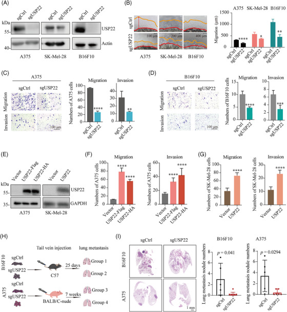
FIGURE 2.

USP22 loss suppresses melanoma metastasis both in vitro and in vivo. (A) Western blot analysis of the indicated proteins in control (sgCtrl) and USP22‐knockout (sgUSP22) A375, SK‐Mel‐28, and B16F10 cells. (B) 3D Matrigel drop invasion assay for A375, SK‐Mel‐28, and B16F10 cells after USP22 knockout (sgUSP22). Indicated medium was exchanged every 3 days. The distances of migrated cells away from edge of the matrigel drop were measured as migration (µm) on Day 6. Experiments were triplicate repeated. Representative images and scare bars are shown. (C and D) Transwell assay quantifying the migration (without extracellular matrix) and invasive (with extracellular matrix) capacity for A375 and B16F10 cells after USP22 knockout (sgUSP22). Migrated and invaded cells were determined for 12−20 h. Five random areas were selected. (E) Western blot analysis of the indicated proteins in control (Vector) and USP22‐overexpressing A375 and SK‐Mel‐28 cells. (F and G) Transwell assay quantifying the migration and invasion for A375 (F) and SK‐Mel‐28 cells (G) after USP22 overexpression. (H and I) Schematic view and representative hematoxylin–eosin (H&E) images of lung metastasis of mice after tail vein injection of B16F10 or A375 cells with USP22‐knockout (sgUSP22) or control (sgCtrl) cells. Two‐tailed unpaired Student's t‐test was performed in (B–D, G, and I), and one‐way ANOVA was used in (F). *p < 0.05, **p < 0.01, ***p < 0.001, ****p < 0.0001.
