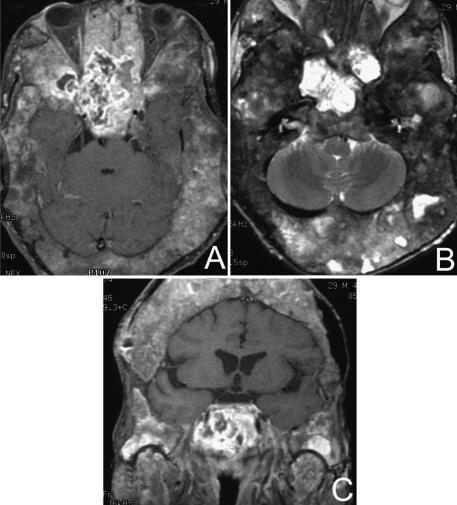
Figure 1.
MRIs of a radiation–induced osteosarcoma in a patient with severe fibrous dysplasia of the skull and skull base. (A) Gadolinium–enhanced, T1–weighted axial image with fat suppression shows a large tumor in the region of the sphenoid and sella. (B) T2–weighted fast spin–echo, axial and (C) gadolinium–enhanced, T1–weighted coronal image with fat suppression of the same lesion.

