
Figure 1D.
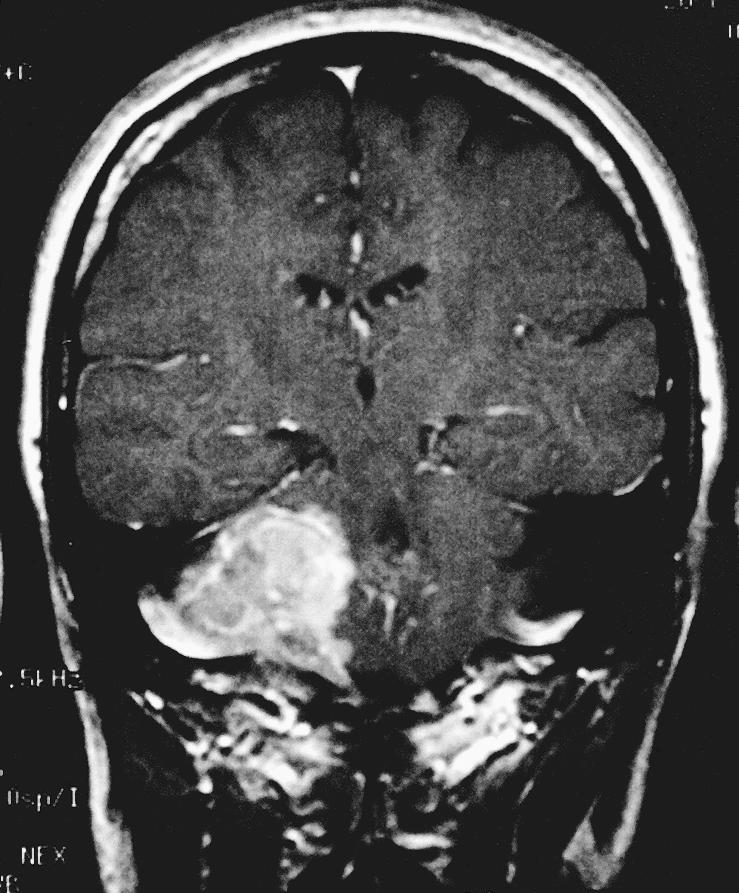
Coronal T1–weighted image with gadolinium show an extra–axial mass of the right CPA. A dural tail extends into the internal auditory canal, jugular foramen, and hypoglossal canal. The lesion, 3.4 × 2.3 × 2.5 cm, exerted mass effect on the midbrain. It was hyperintense on T1–weighted images and isointense on T2–weighted images with a homogeneous appearance. After gadolinium administration it enhanced prominently. A second smaller dural–based lesion was found in the lateral aspect of the right posterior fossa, invaginating into the adjacent cerebellum.