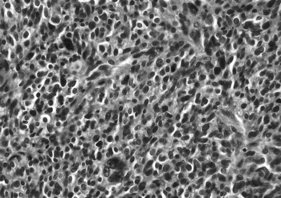
Figure 2B.
Hematoxylin and eosin histological sections seen at magnification 40×. Microscopic examination showed a tumor infiltrating into the surrounding connective tissue. The nuclei exhibited pleomorphism and hyperchromasia. Brown pigment was seen within the tumor, but mitoses were difficult to find. A CPA melanoma was diagnosed.

