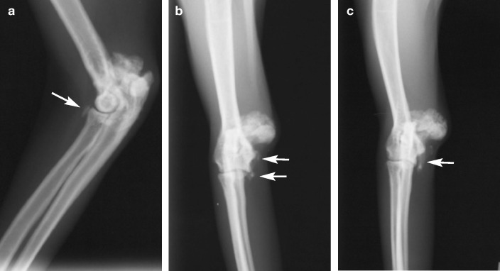
FIG 1.
Mediolateral (a), craniocaudal (b) and craniocaudal oblique (c) radiographs of the left elbow of an elderly cat with left forelimb lameness. There is a well-circumscribed mineralised lesion adjacent to the medial epicondylar ridge of the left humerus. There is also a conical-shaped bony structure (arrow; c) extending from the joint to the proliferative mass, which was speculated to be a mineralised ‘outpocketing’ of synovial membrane. There is no lytic component to the lesion, or the adjacent humerus. As well as these lesions, there is evidence of chronic osteoarthritis of the left elbow (eg, the supinator sesamoid bone [arrow; a] adjacent to the radius is more obvious than normal), increased radiodensity of the subchrondral bone in the ulna (a), and mineralised structures on the medial humeral epicondyle and medial extent of the articular surface (arrows; b), which could be enthesiophytes, further osteochondromas or mineralised free bodies

