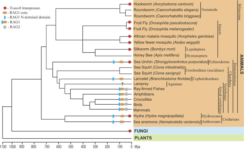
Figure 1. Schematic Presentation of Transib transposons, RAG1, RAG2, and RAG1-Like Proteins in Eukaryotes.
The basic timescale of the evolutionary tree is based on published literature [49–51]. Red circles mark species in which Transib TPases were found. Gray squares indicate RAG2; orange and blue ellipses show the RAG1 core and RAG1 N-terminal domain, respectively. Overall taxonomy, including common and Latin names, is reported on the right side of the figure. A question mark at the lamprey lineage indicates insufficient sequence data. A lack of any labels means that the Transib TPase and RAG1/2 are not present in the sequenced portions of the corresponding genomes. Among branches lacking Transib TPases, only lamprey and crocodile genomes are not extensively sequenced to date. In sea anemone, the RAG1 core–like protein is capped by the ring finger motif, which also forms the C-terminus in the RAG1 N-terminal domain. In fungi, the Transib TPase was detected in soybean rust only.

