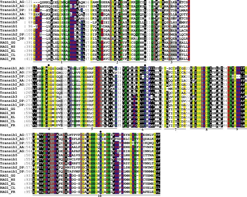
Figure 3. Multiple Alignment of Ten Conserved Motifs in the RAG1 Core Proteins and Transib TPases.
The motifs are underlined and numbered from 1 to 10. Starting positions of the motifs immediately follow the corresponding protein names. Distances between the motifs are indicated in numbers of aa residues. Black circles denote conserved residues that form the RAG1/Transib catalytic DDE triad. The RAG1 proteins are as follows: RAG1_XL (GenBank GI no. 2501723, Xenopus laevis, frog), RAG1_HS (4557841, Homo sapiens, human), RAG1_GG (131826, Gallus gallus, chicken), RAG1_CL (1470117, Carcharhinus leucas, bull shark), RAG1_FR (4426834, Fugu rubripes, fugu fish). Coloring scheme [43] reflects physiochemical properties of amino acids: black shading marks hydrophobic residues, blue indicates charged (white font), positively charged (red font), and negatively charged (green font); red indicates proline (blue font) and glycine (green font); gray indicates aliphatic (red font) and aromatic (blue font); green indicates polar (black font) and amphoteric (red font); and yellow indicates tiny (blue font) and small (green font). The species abbreviations for the Transib transposons are as follows: AA, yellow fever mosquito; AG, African malaria mosquito; DP, D. pseudoobscura fruit fly. (Transib1 through Transib5 are from the fruitfly D. melanogaster).

