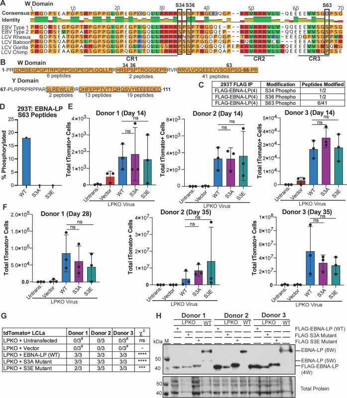
Fig 2. Phosphorylation of EBNA-LP is not required for EBV-infected naïve B cell transformation.
A. Known phosphorylated serines in EBNA-LP W domain across human EBV strains (Type 1 is B95-8, Type 2 is AG876) and EBNA-LP homologs in primate LCV strains. Identity bar indicates conservation of amino acids, with green indicating highly conserved regions, yellow indicating less conserved, and red are poorly conserved regions. Colors of residues indicate amino acid properties. Phosphorylated serines are indicated by black boxes. Previously defined conserved regions (CR1, CR2, CR3) are underlined (22). B. Amino acid sequence coverage of FLAG-tagged EBNA-LP (wild type) construct encoding 4 W domains expressed in 293T cells, followed by mass spectrometry of FLAG immunoprecipitation. Underlined and highlighted residues indicate peptides identified by mass spectrometry, with the number of peptides indicated below. Phosphorylated residues are red, with residue number above. C. Abundance of phosphorylated peptides for all identified phospho-serines out of the total peptides spanning that residue from FLAG immunoprecipitation in B. D. Average percent of total Tandem Mass Tag (TMT) signal for peptides with S63 phosphorylation when FLAG-tagged EBNA-LP (wild type), the EBNA-LP S3A mutant, and EBNA-LP S3E mutant are expressed in 293T cells prior to FLAG immunoprecipitation, TMT labeling, and mass spectrometry (n = 3). E. Total tdTomato positive cells at 14 days post infection for each donor in LPKO virus infected, trans-complemented cells (n = 3). Significance determined by unpaired t-test. Mean and standard deviation are plotted. F. Total tdTomato positive cells at 28 or 35 days weeks post infection for each donor (n = 3). G. Total tdTomato+ LCLs generated per condition. #Indicates conditions in which tdTomato negative LCLs were generated. P values calculated using Fisher’s exact test to compare transformation outcomes to LPKO + Vector. **** indicates a p value < 0.0001. *** indicates a p value < 0.001. H. Trans-complemented LPKO LCLs express wild type and mutant FLAG-tagged EBNA-LP. W indicates number of W domains in EBNA-LP protein expressed from virus or expression construct. M indicates molecular weight marker.

