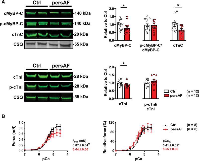
Figure 3.
Myofilament protein expression and contractile response to cytosolic Ca2+ in control and persAF. A, Immunoblots (upper left) and quantification (upper right) of cMyBP-C (cardiac myosin binding protein-C), its phosphorylated state (P-cMyBP-C), and cTnC (cardiac troponin-C) in atrial samples from controls and patients with persAF, normalized to CSQ (calsequestrin), except for P-cMyBP-C, which was normalized to total cMyBP-C. Immunoblots (lower left) and quantification (lower right) of cTnI (cardiac troponin I) and its phosphorylated state (P-cTnI) in atrial samples from controls and patients with persAF, normalized to CSQ and total cTnI, respectively. B, Absolute (left) and normalized (right) force-pCa relationship of skinned muscle fibers of controls and patients with persAF with mean±SEM of maximum force (Fmax) and calcium sensitivity (pCa50). *P<0.05 vs control. n=number of patients. Normality of data was determined by Shapiro-Wilk test, whereas comparison was made using the Student t test with Welch correction. Ctrl indicates control; and persAF, persistent atrial fibrillation.

