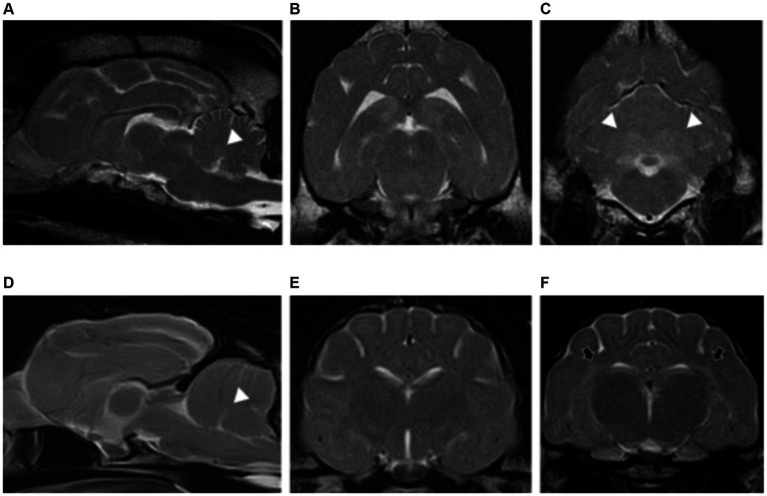
Figure 10.
Sagittal (A) and transverse plane T2-weighted images at the level of the forebrain (B) and cerebellum (C) of a dog with GM2 gangliosidosis. Sagittal (D) and transverse T2-weighted of the forebrain (E,F) of a cat with GM1 gangliosidosis. Similar findings are visible for both patients. There is diffuse hyperintensity of the white matter of the forebrain with subsequent decrease of the normal grey matter/white matter definition. In the cat, this is especially marked at the corona radiata (short black arrows) (F). The changes are also visible affecting the cerebellar white matter of both patients (white arrowheads) (A,C,D).

