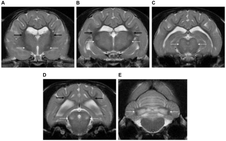
Figure 13.
Transverse plane T2-weighted images of the brain of a cat with L-2-hydroxyglutaric aciduria. Note the diffuse cortical grey matter swelling of the forebrain and cerebellum with increased signal intensity (A–E). There are bilateral and symmetrical lesions affecting the globus pallidus and putamen (long white arrows) (A), thalamus (long white arrow) (B), oculomotor nuclei (long white arrows) (C), lateral lemniscus (long white arrows) (D) and cerebellar nuclei (long white arrows) (E). The subcortical white matter is also mildly affected (long black arrows).

