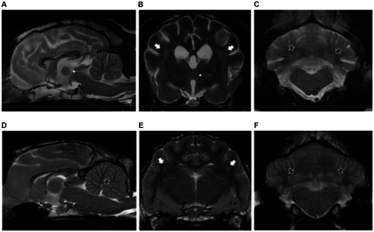
Figure 2.
Sagittal and transverse plane T2-weighted images at the level of the forebrain and cerebellum of a dog (A–C) and cat (D–F) diagnosed with hepatic encephalopathy. Both patients have bilateral and symmetric hyperintensities affecting the centrum semiovale and extending towards the corona radiata (short white arrows) (B,E) as well as at the cerebellar nuclei (short black arrows) (C,D,F). Note also the moderate brain atrophy in the canine patient evidenced by the decreased interthalamic height (long white arrows) and widening of the cerebral and cerebellar sulci (A–C).

