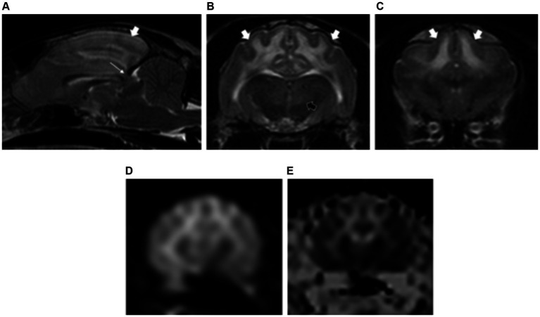
Figure 7.
Sagittal T2-weighted image (A) of a cat with hypertensive encephalopathy. Note the associated mass effect with caudal displacement of the midbrain across the tentorium cerebelli (caudal transtentorial herniation) (long white arrow). Transverse plane T2-weighted images showing the white matter hyperintensities (short white arrows) extending from the occipital and parietal lobes (A,B) to the frontal lobes (C). Bilateral and symmetric hyperintensities are also noted affecting the thalamus (short black arrow) (B). Diffusion-weighted (D) and apparent diffusion coefficient (ADC) map images (E) showing facilitated diffusion of the white matter lesions, confirming the vasogenic origin of the oedema.

