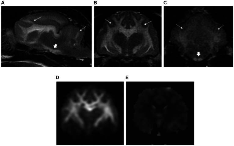
Figure 8.
Left parasagittal (A) and transverse plane T2-weighted images at the level of the forebrain (B) and cerebellum (C) of a cat with bromethalin intoxication. There is severe brain swelling with effacement of the normal cerebral and cerebellar sulci diffusely. Severe diffuse hyperintensities are visible along the white matter of the forebrain (long white arrows), cerebellum (dashed long white arrows) and along the crus cerebri towards the pyramidal tracts of the medulla oblongata (short white arrows). Diffusion-weighted (D) and apparent diffusion coefficient (ADC) map images (E) showed restricted diffusion of the white matter lesions, confirming the cytotoxic origin of the oedema.

