
Fig. 2.
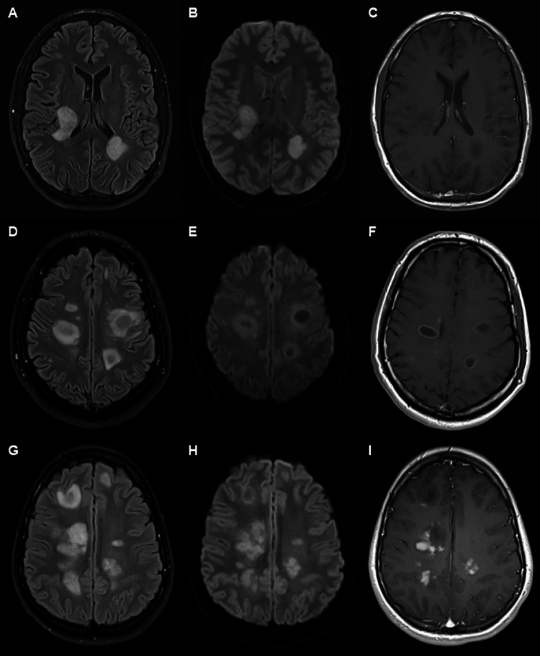
Three patterns of lesion in ADEM-like lesions. Vertical panels show FLAIR sequences (right), diffusion weighted images (centre) and T1 with contrast (left). Horizontal panels show individual cases. Upper panel (A–C) shows case with FLAIR and DWI hyperintensity with T1 hypointensity, but no Gd-enhancement. Middle panel (D–F) shows ring pattern hyperintensity on FLAIR and DWI with central hypointensity and ring-enhancement on T1 sequence. Lower panel (G–I) shows predominantly heterogeneous FLAIR and DWI hyperintensity with heterogeneous Gd-enhancement. There are additional lesions showing central T1 hypointensity and no enhancement.