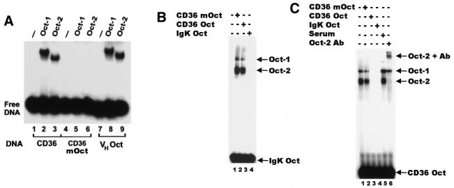
Figure 2.
Specific binding of Oct-1 and Oct-2 to the CD36 octamer. (A) Gel retardation analysis of in vitro translated Oct-1 and Oct-2, demonstrating specific binding to the octamer site within the CD36 promoter. Reactions were performed with either the wild-type CD36 probe (CD36, lanes 1–3), the mutant octamer CD36 probe (CD36mOct, lanes 4–6) or a known octamer element (VHOct, lanes 7–9). Reticulocyte lysates were programmed with cDNAs for the proteins indicated above each lane. Lanes 1, 4 and 7 contain unprogrammed reticulocyte lysates. (B) Gel retardation analysis showing the CD36 octamer (CD36 Oct) specifically competes with the IgK octamer (IgK Oct). Nuclear extracts from WEHI231 cells were incubated with radiolabelled IgK Oct in the presence of 100-fold molar excess of competitor DNA as indicated above the lanes. The known Oct-1 and Oct-2 complexes are indicated. (C) Oct-2 specifically binds to the CD36 octamer. Nuclear extracts from WEHI231 cells were incubated with radiolabelled CD36 Oct in the presence of 100-fold molar excess of competitor DNA as indicated above the lanes (lanes 2–4). Lanes 5 and 6 contain serum and Oct-2-specific antibody, respectively. The identity of each of the complexes is indicated.

