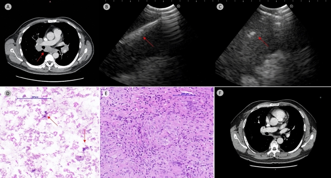
Figure 1.
Representative case. Contrast enhanced computed tomography show bilateral hilar and mediastinal lymphadenopathy (arrow, Panel A). EBUS-TBNA was performed with 22-gauge TBNA needle with a total of 5 passes (arrow, Panel B). 1.1 mm cryoprobe was then inserted after mucosal incision with high-frequency needle knife (arrow, Panel B); a total of 5 cryo-passes was performed with total freezing time of 22 s (average 4.4 s freezing time per cryo-pass). TBNA cell block revealed only cellular debris with scanty macrophages (arrow, hematoxylin & eosin stain, Panel D). Collectively, the TBMC specimen measured 11 mm in total aggregate diameter which demonstrated a well-formed non-caseating granuloma consistent with clinical suspicion of sarcoidosis. (hematoxylin & eosin stain, Panel E). Post treatment with steroid and methotrexate demonstrate complete resolution of hilar and mediastinal lymphadenopathy (Panel F).

