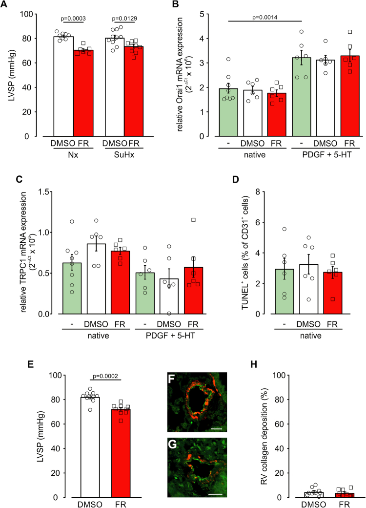
Figure EV5. FR effects on Hx-induced PH in vivo and mPASMCs as well as mLECs in vitro.
(A) Statistical analysis of LVSP in mice treated with the solvent DMSO or FR (10 µg/mouse i.p., Monday to Friday) during exposure to Nx (21% O2, DMSO: n = 8, FR: n = 7) or SuHx (10% O2, DMSO: n = 10, FR: n = 10) for 3 weeks. (B, C) Statistical analysis of relative Orai1 (B) and TRPC1 (C) mRNA expression in native mPASMCs (n = 8) and mPASMCs treated with solvent DMSO or FR (10−6 M) with or without additional PDGF (40 ng/ml) + 5-HT (10−6 M) stimulation for 12 h, each n = 6 normalized to 18 S housekeeping gene, ns indicate different wells derived from at least two different passages. (D) Amount of TUNEL+ CD31+ mLECs after 2 days without treatment (n = 6) or with DMSO (n = 6) or FR (10−6 M, n = 6) treatment. (E) Statistical analysis of LVSP in mice treated with the solvent DMSO (n = 9) or FR (10 µg/mouse i.p., Monday to Friday, n = 9) in the last 2 weeks of 5 weeks SuHx exposure. (F, G) vWF/α-SMAC staining of PAs in lung sections from SuHx-DMSO (F) and SuHx-FR-treated mice (G), scale bars: 20 µm. (H) Statistical analysis of collagen deposition in the right ventricle in mice treated with the solvent DMSO (n = 9) or FR (10 µg/mouse i.p., Monday to Friday, n = 9) in the last 2 weeks of 5 weeks SuHx exposure. Data information: Values are expressed as mean ± SEM. (A–D) One-way ANOVA, Tukey’s post hoc test, (E, H) Unpaired student’s t-test. Source data are available online for this figure.

