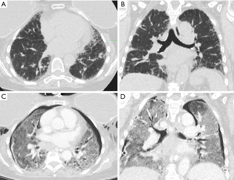
Figure 1.
Lung parenchyma pre- and post-ILD exacerbation. (A,B) Show axial and coronal views respectively of CT chest prior to cryobiopsy demonstrating unspecified ILD. (C,D) Show axial and coronal views at similar levels 24 hours post cryobiopsy with small bilateral pneumothoraces with diffuse ground glass infiltrates consistent with AE-ILD. CT, computed tomography; ILD, interstitial lung disease; AE-ILD, acute exacerbation of interstitial lung disease.

