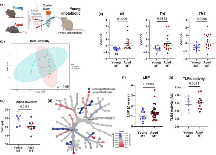
FIGURE 3.

Aged microbiota transplantation (MT) into germ‐free mice partially recapitulates colon inflammation and signs of barrier disruption. (a) A schematic representation of the gnotobiotic experiment. A mixture of intestinal contents (cecal and colonic contents, and feces) from young (3–4 months) or aged (18–20 months) CONV‐R donor mice was transferred to young germ‐free recipient mice. All measurements were performed in young MT and aged MT samples, 4 weeks after colonization. (b) β‐diversity analysis: Principal coordinate analysis plot using the Bray–Curtis index from young MT (red spheres) and aged MT (blue spheres) mice. Dotted line ellipses correspond to clusters in each group. (c) α‐diversity analysis: index Faith‐pd represents community richness. (d) Heat tree plotting of colonic taxa abundance (aged MT vs. young MT) to allow quantitative visualization of community diversity data comparing mice that received microbiota from the two age groups. (e) Colonic inflammatory gene expression of recipient gnotobiotic mice was assessed by qPCR. (f) Lipopolysaccharide‐binding protein (LBP) in serum, analyzed by ELISA kit according to manufacturer's recommendations. (g) TLR4 activity in colon contents, assessed using human embryonic kidney (HEK)‐blue‐mTLR4. Relative fluorescence intensity was used as an estimation for TLR4 activity, considering gnotobiotic mice plus young MT as control (Relative AU =1). Data correspond to n = 3 cohorts with n = 10–15 per group.
