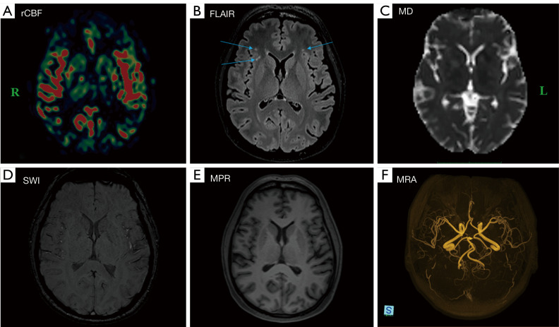
Figure 6.
A representative map (A) of normal rCBF in a 47-year-old symptomatic female patient is provided, along with the corresponding results from complementary MR images (B-F). The arrows indicate hyperintense lesions observed following a history of COVID-19 infection. Apart from the lesions identified on 3D T2-FLAIR images (B), there are no abnormal findings noted on MD, SWI, MPR gradient echo single-shot echo-planar imaging sequence, or MRA images (C-F). L indicates left, and R indicates right. rCBF, regional cerebral blood flow; FLAIR, fluid attenuated inversion recovery; MD, mean diffusivity; SWI, susceptibility-weighted imaging; MPR, magnetization prepared rapid; MRA, MR angiography; MR, magnetic resonance; COVID-19, coronavirus disease 2019; 3D, three-dimensional.

