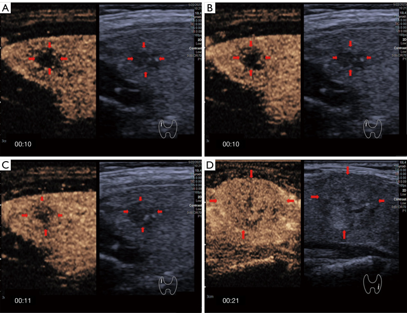
Figure 4.
Enhancement direction on CEUS. (A-C) Images showing centripetal enhancement of the same PTC nodule (red arrows) in the right lobe of thyroid gland of a 69-year-old woman. (D) Image of a case of a benign nodule (red arrows) in the left lobe of thyroid gland of a 41-year-old woman showing a feature of scattered enhancement on CEUS. The minutes and seconds after the contrast media injection are indicated by numbers in the bottom left corner of each panel. CEUS, contrast-enhanced ultrasound; PTC, papillary thyroid carcinoma.

