Figure 6.
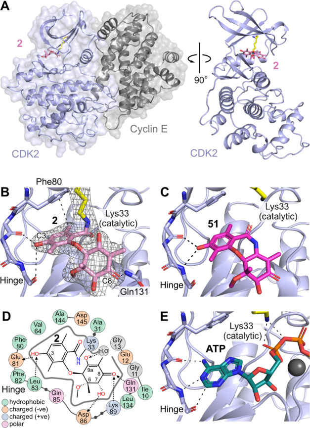
(A) Co-crystal structure of CDK2/cyclin E in complex with 2 (PDB ID 9BJB). (B) Active site of CDK2 with bound 2, showing H-bonding interactions from C3-OH to the kinase hinge; electron density map of 2 overlaid in mesh (2fo-fc contoured at 1 sigma) demonstrates covalent attachment to Lys33. (C) Binding of amide analog XC208 51 (PDB ID 9BJC) without covalent interaction, demonstrating a prereaction complex. (D) 2D map of binding site interactions between CDK2 and 2, showing covalent (solid line) and H-bonding (dotted lines) positions. (E) Structure of CDK2/cyclin A with ATP bound showing similar interactions to 2 and 51 at the hinge and catalytic Lys33 (PDB ID 4EOQ).

