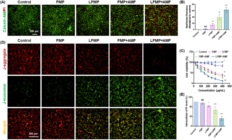
Fig. 3.
A Live/dead cell staining assay of the EMT-6 cells after treatment with different groups (scale bar = 200 µm). B Statistical analysis of red/green fluorescence in A by flow cytometry. C The cytotoxicity of EMT-6 cells with different treatments. D Determination of the population of polarized/depolarized mitochondria using JC-1. In apoptotic cells, JC-1 exists in the monomeric form because of the low mitochondrial membrane potential, staining the cytosol green. In live nonapoptotic cells, JC-1 accumulates as aggregates in the mitochondrial membrane which stains red. E Concentrations of intracellular ATP using an ATP Determination Kit. Data are shown mean ± SD, n = 5. *p < 0.05, **p < 0.01, ***p < 0.005

